Revolutionizing Cancer Diagnosis with AI Pathologists
Written on
Chapter 1: The Need for Speed in Cancer Diagnosis
In the realm of cancer surgery, having quick and precise information about the tissue being operated on is vital for guiding the surgeon’s next steps. Traditionally, this information is obtained through pathological analysis, which requires examining tissue samples under a microscope to identify any abnormalities or signs of cancer.
This conventional approach is often time-consuming and depends heavily on the expertise of trained pathologists for accurate interpretation. Consequently, patients may find themselves returning to the hospital days later for further surgery, causing stress for both them and their families while also increasing healthcare expenses. Fortunately, researchers at the Max Planck Institute for the Science of Light (MPL) in Erlangen have devised a groundbreaking method that could change how tissue samples from cancer patients are analyzed.
The team, in collaboration with esteemed institutions like the Max-Planck-Zentrum für Physik und Medizin (MPZPM), Friedrich-Alexander-Universität Erlangen-Nürnberg, University Hospital Erlangen, and Fraunhofer Institute for Process Automation (IPA) in Mannheim, has developed an artificial pathologist that utilizes artificial intelligence to interpret data generated by their innovative technique.
“This approach allows us to conduct a physical examination of individual cells, offering significantly more information than traditional methods,” says Dr. Markéta Kubánková, the lead researcher.

Chapter 1.1: A New Era of Tissue Analysis
The research team, led by Dr. Despina Soteriou, recently published findings that demonstrate their method's capability to quickly and accurately identify tumor tissue in samples. They envision a future where this technology could assist or even replace conventional pathological analysis.
The novel method combines laser microdissection with mass spectrometry imaging to scrutinize cells in cancer patient tissue samples.

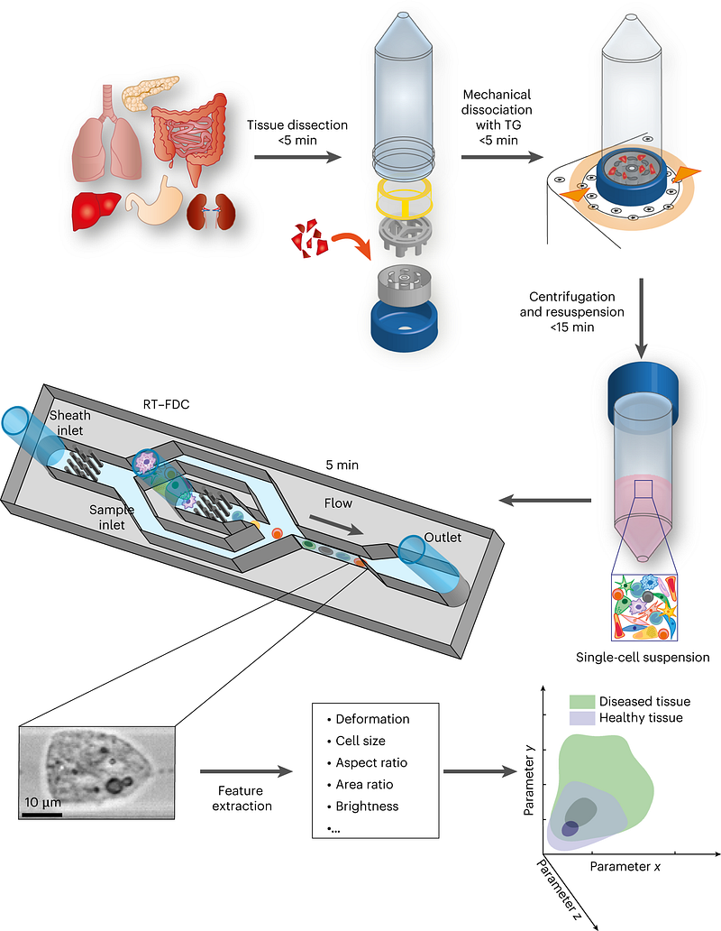
The biopsy process involves a tissue grinder that breaks down the sample for real-time deformability cytometry (RT-DC) analysis. Using a laser, specific cells are extracted, which are then analyzed through mass spectrometry imaging. An artificial pathologist interprets this data using machine learning algorithms, enabling clinicians to swiftly and accurately detect abnormalities or cancer indicators without needing a trained pathologist.
Next, the researchers tested this groundbreaking method on mice tissue, using a specialized device that processes samples into individual cells in just five minutes. These cells are analyzed in real-time using a combination of fluorescence and deformability cytometry (RT-FDC), which employs a syringe pump to pass single cells through a microscopic constriction, exposing them to hydrodynamic shear stress.
Video Description: This video explores the integration of molecular diagnostics and machine learning in diagnosing and discovering therapies for brain tumors.
Chapter 1.2: Understanding Cell Deformability
Cell deformability plays a crucial role in assessing the invasiveness of cancer cells and diagnosing diseases. More deformable cancer cells are often more invasive, leading to metastasis. The RT-FDC technique can analyze up to 1,000 cells per second—36,000 times faster than older methods. As cells navigate through the microscopic constriction, RT-FDC captures images to evaluate their physical characteristics and distinguish tissue cell subtypes through image analysis alone.
This innovative technology presents multiple advantages over traditional pathological analysis. First, it drastically reduces analysis time to just 30 minutes for accurate solid tumor evaluations. Second, it eliminates the need for a trained pathologist, thereby lowering healthcare costs and improving access to cancer diagnoses. Ultimately, this method could aid clinicians in assessing disease severity or differentiating between various types of inflammatory bowel disease. Clinical trials will be the next step in testing this technology.

Chapter 2: Future Directions in Cancer Diagnosis
With the promising advancements in AI-driven pathological analysis, researchers are optimistic about the future of cancer diagnosis and treatment.
Video Description: This video discusses the future of computational pathology and AI innovations at Mayo Clinic, highlighting transformative approaches in healthcare.
The complete findings of this groundbreaking research were published in the Journal of Nature Biomedical Engineering.
Stay updated with further insights and developments by following my work on Medium.